
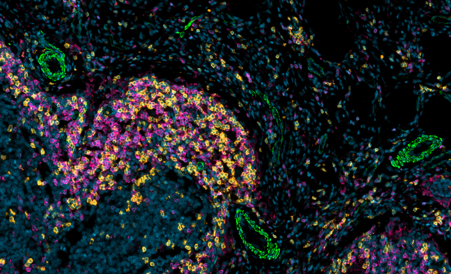
Cyclic Immunofluorescence (CycIF) & Spatial Biology Services
High-plex multiplex immunofluorescence and spatial biology workflows for tumor microenvironment analysis, immune profiling, biomarker discovery, and translational research powered by Lunaphore COMET™ technology.
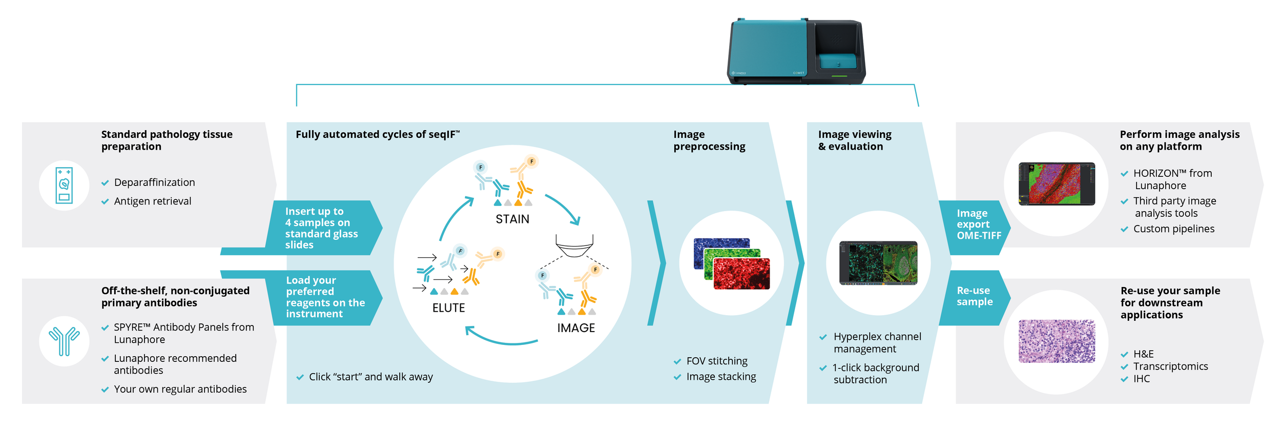
Lunaphore COMET™ Multiplex Tissue Imaging
Automated cyclic immunofluorescence workflows for scalable high-dimensional tissue imaging and spatial biomarker analysis.
What is Cyclic Immunofluorescence (CyCIF)?
CyCIF enables repeated cycles of antibody staining and fluorescence imaging to visualize dozens of protein biomarkers within intact tissue architecture while preserving spatial context and cellular organization.
Multiplex Immunofluorescence & Spatial Biology Services:
IMCO provides end-to-end CyCIF services including experimental design consultation, antibody panel optimization, tissue imaging, spatial analysis, and downstream bioinformatics support.
Why we use COMET cycIF in IMCO labs:
Ultra-high plexing: simultaneous visualization of 30+ markers in a single tissue section with precise spatial resolution.
Same slide H&E for AI driven data analysis.
Spatial biology, uncoupled from single-marker limitations: understand cell–cell interactions, neighborhoods, and microenvironment patterns.
FFPE-compatible workflows: work with routinely collected clinical samples without complex preprocessing.
Rapid, automated workflow: streamlined protocols reduce hands-on time and improve consistency.
Quantitative, reproducible data: standardized cycles enable robust image analysis and cross-sample comparability.
End-to-end readiness: integrates with our imaging platforms and data analytics to accelerate discovery and decision-making. Learn more about the Lunaphore Comet here.

Featured Panels
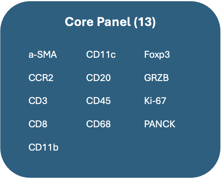
Core Panel
Targets: CD8 T Cells, Tregs, Th Cells, B Cells, Monocytes, Macrophages, DCs, Neoplastic Cells, Fibroblast-like Cells
Function: Proliferation, Cytotoxicity
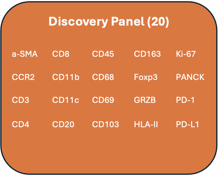
Discovery Panel
Targets: CD8 T Cells, Tregs, Th Cells, Unconventional T Cells, B Cells, Monocytes, Macrophages, DCs, Other APCs, Neoplastic Cells, Fibroblast-like Cells
Function: Proliferation, Activation, Checkpoint, Cytotoxicity, Tissue Residency, Anti-inflammatory(M2-like)
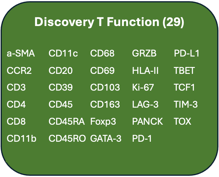
Discovery T-Function
Targets: CD8 T Cells, T-regs, Th1 Cells, Th2 Cells, Unconventional Cells, B Cells, Monocytes, Macrophages, DCs, Other APCs, Neoplastic Cells, Fibroblast-like Cells
Function: Proliferation, Activation, Cytotoxicity, Tissue Residency, Memory, Exhaustion, Self-renewal, Immunosuppression, Anti-inflammatory (M2-like)
Custom Panels
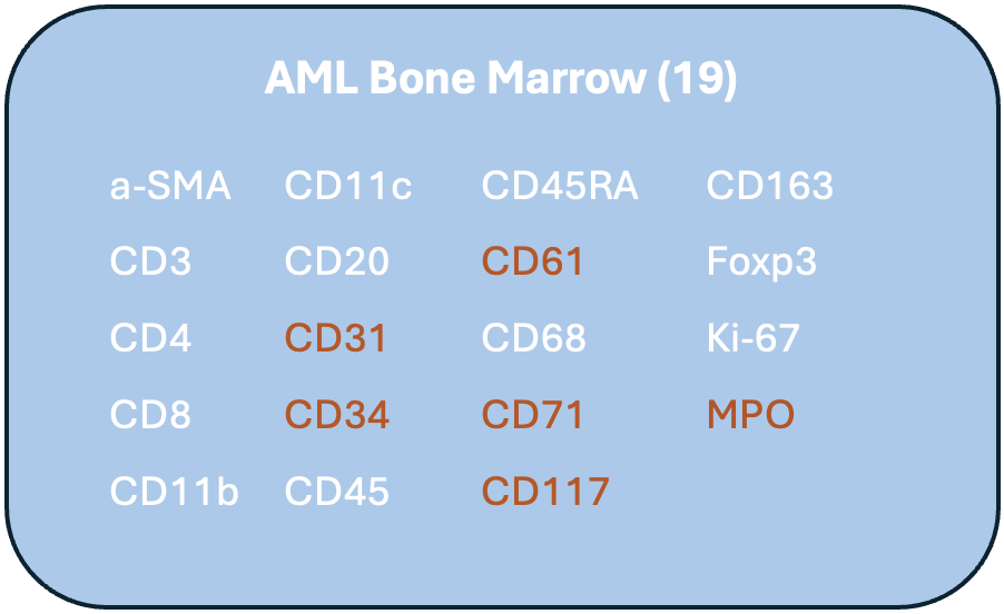
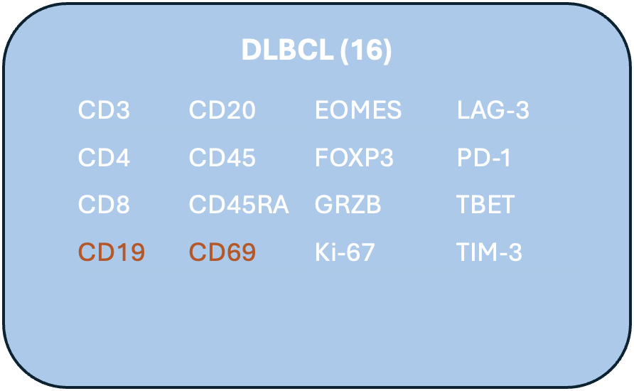
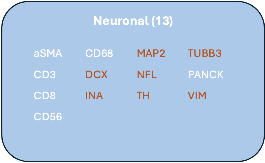
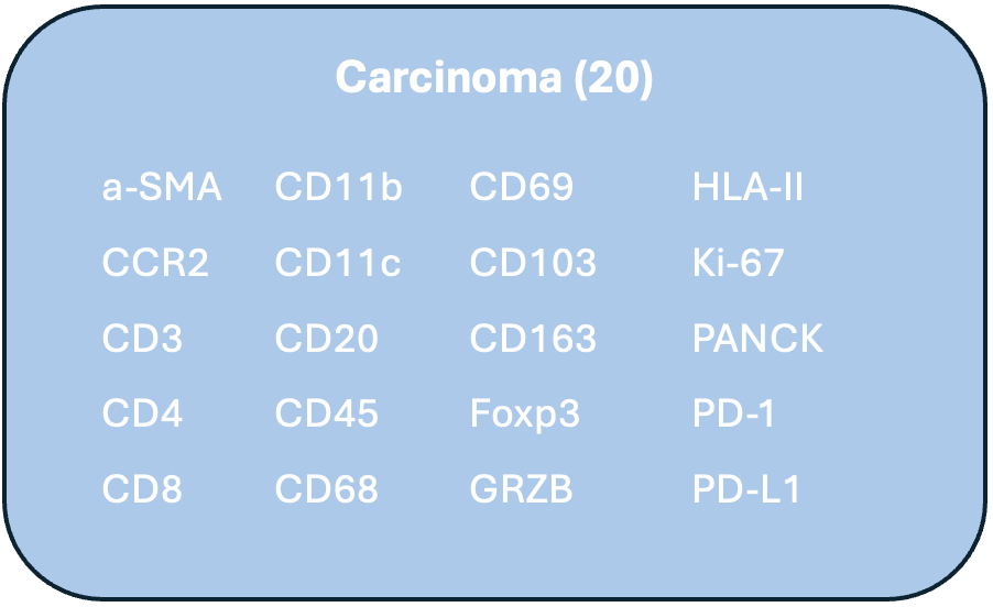
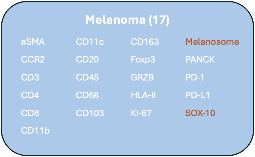
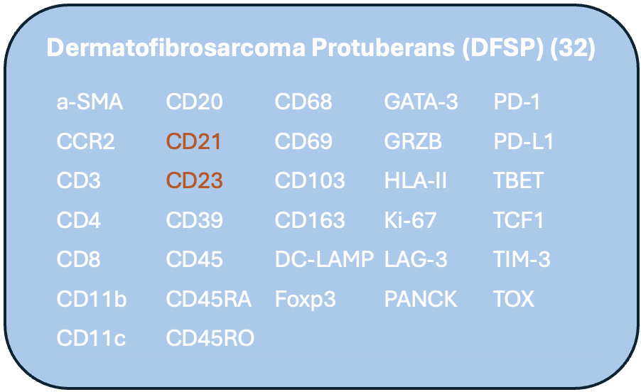
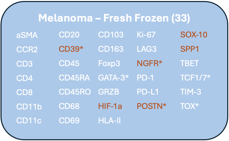
Lunaphore at work
-
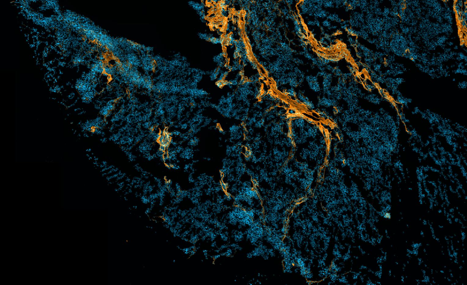
![A digital, neon-like abstract art piece featuring a human face outline in orange lines intertwined with blue digital particles on a black background.]()
Melanoma: DAPI, POSTN
-
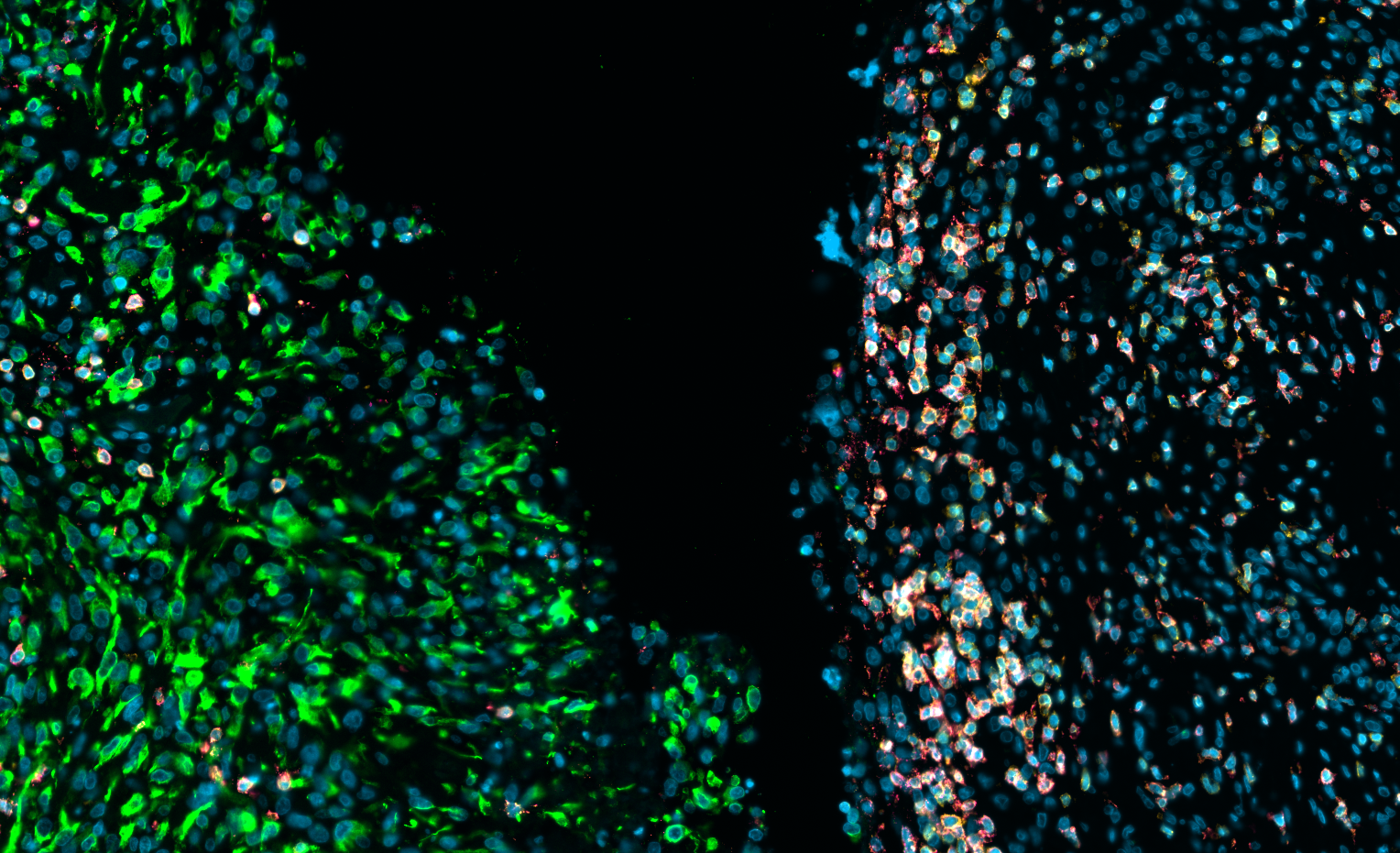
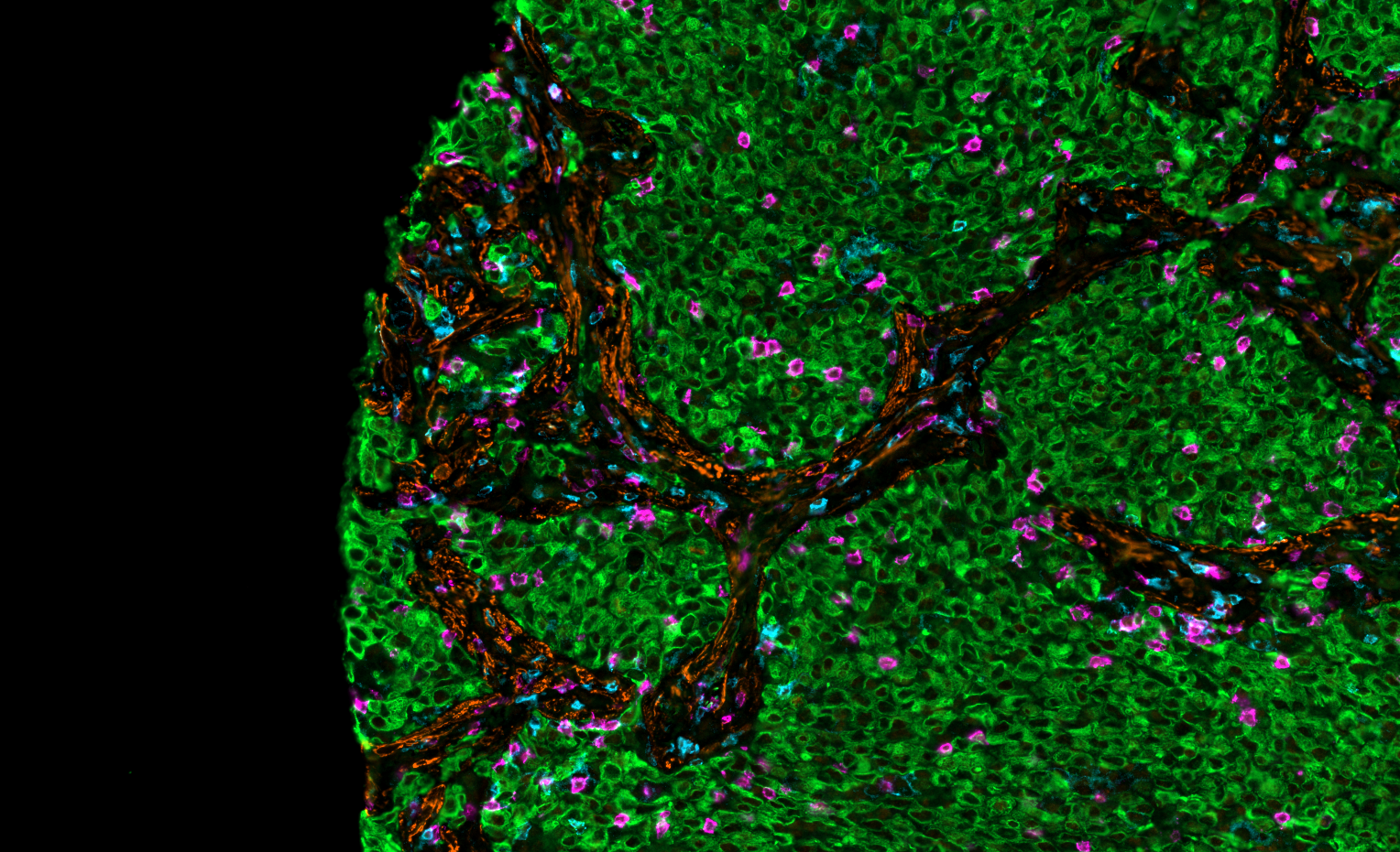
![Close-up of cells stained with fluorescent antibodies, showing green fluorescence on the left and multicolored fluorescence on the right, used for scientific research.]()
Sarcoma: CD3, CD8, Pan-CK
-
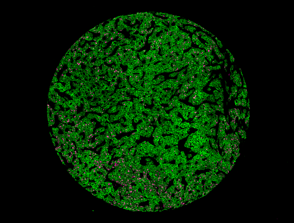
![Breast Cancer: Pan-CK, CD45]()
Breast Cancer: Pan-CK, CD45
-
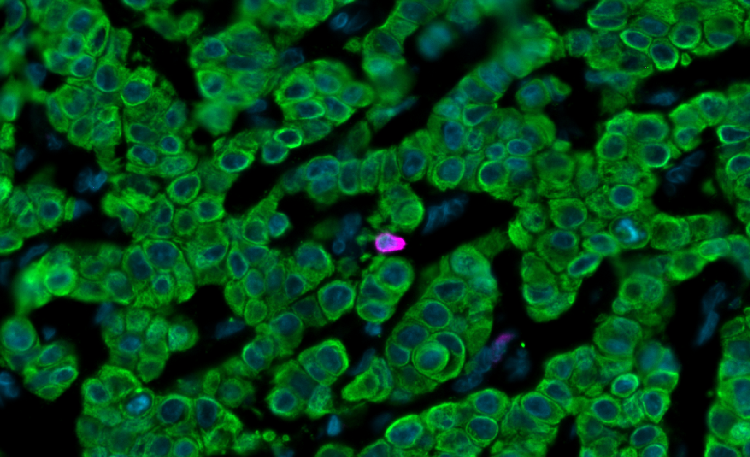
![Breast Cancer: Pan-CK, CD45]()
Breast Cancer: Pan-CK, CD45
-
![Melanoma TLS: CD8, ASMA]()
Melanoma TLS: CD8, ASMA
-
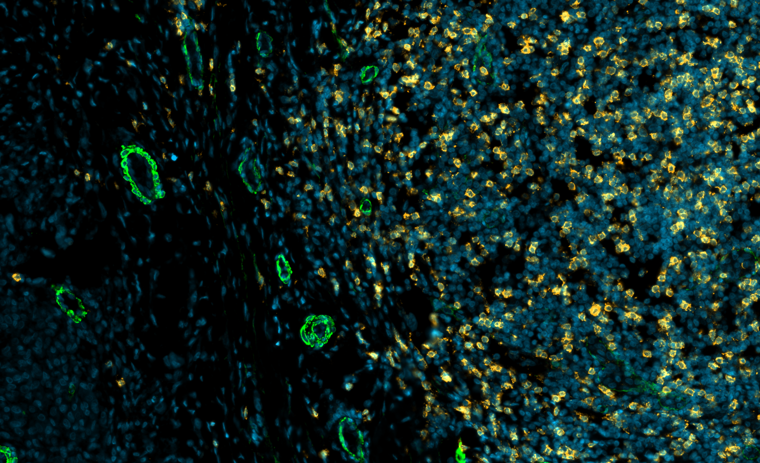
![Lung: CD4, CD8, ASMA, Pan-CK]()
Lung: CD4, CD8, ASMA, Pan-CK
-
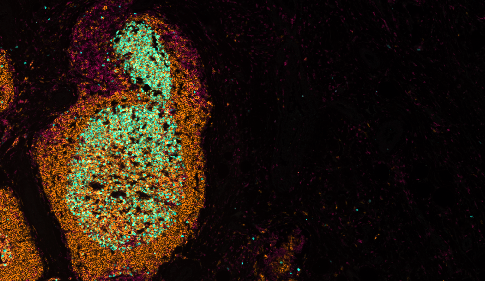
![Melanoma TLS: Ki-67]()
Melanoma TLS: Ki-67
FAQs
-
Cyclic immunofluorescence (CyCIF) is a high-plex imaging technique that uses repeated cycles of antibody staining, fluorescence imaging, and signal removal to visualize dozens of protein biomarkers within a single tissue section. This approach preserves tissue architecture and spatial context, enabling comprehensive characterization of cellular phenotypes and tissue organization.
-
Traditional immunofluorescence is typically limited to a small number of biomarkers due to spectral overlap between fluorophores. CyCIF overcomes this limitation by performing multiple staining and imaging cycles, allowing significantly more biomarkers to be analyzed within the same tissue section.
-
CyCIF is commonly performed on formalin-fixed paraffin-embedded (FFPE) tissue and can also be applied to other validated tissue types depending on study requirements. IMCO works with investigators to determine the most appropriate sample preparation and workflow for each project.
-
CyCIF is widely used for tumor microenvironment characterization, immune profiling, spatial biology, biomarker discovery, translational pathology, neuroinflammation research, and studies requiring high-dimensional tissue imaging while preserving spatial relationships between cells.
-
The Lunaphore COMET™ is a fully automated cyclic immunofluorescence platform that enables high-plex multiplex tissue imaging through iterative staining and imaging cycles. It delivers reproducible, scalable spatial biology workflows for translational research and biomarker discovery.
-
The number of biomarkers depends on the antibody panel design and study objectives. CyCIF supports high-plex imaging workflows capable of measuring dozens of protein biomarkers within a single tissue section while maintaining spatial context. We commonly operate panels of over 30 markers in a single run.
-
Yes. In addition to tissue staining and imaging, IMCO provides downstream spatial analysis services including single-cell segmentation, cell phenotyping, neighborhood analysis, biomarker co-expression analysis, tissue architecture mapping, and publication-ready data visualization.
-
Yes. CyCIF data can be integrated with complementary datasets such as Olink® proteomics, flow cytometry, mass cytometry (CyTOF), genomics, and transcriptomics to provide a more comprehensive understanding of biological systems and disease mechanisms.
-
Unlike conventional IHC, which typically evaluates one or a few markers per tissue section, CyCIF enables simultaneous analysis of dozens of biomarkers while preserving spatial context. This provides deeper insights into cell populations, cellular interactions, and tissue organization.
-
Our team collaborates with investigators throughout the project lifecycle, providing guidance on study design, antibody panel selection, tissue preparation, imaging workflows, quality control, spatial analysis, and biological interpretation to ensure data are aligned with each study's scientific objectives.