Bioinformatics & Research Analytics Services
Complex multi-omics datasets become actionable biological insights through integrated bioinformatics, statistical analysis, biomarker discovery, and data visualization services. IMCO supports Olink proteomics, CycIF spatial biology, Mass cytometry, genomics, and translational research programs with end-to-end analytical expertise.
Click here to download our sample report!

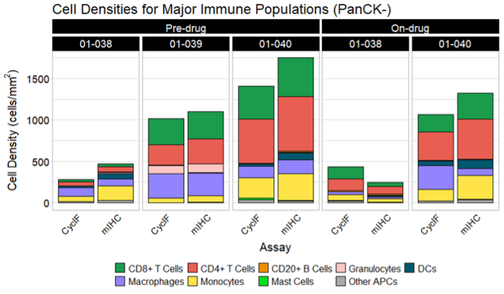
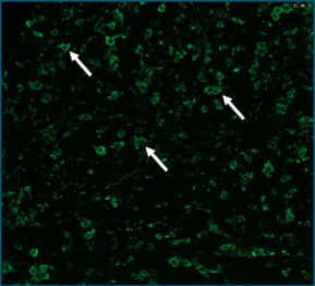
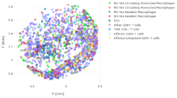
Spatial Biology & Cyclic ImmunoFluorescence Analysis
Transform high-dimensional spatial imaging datasets into biologically meaningful insights through cell segmentation, spatial phenotyping, neighborhood analysis, tissue architecture mapping, and tumor microenvironment characterization.
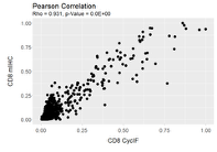
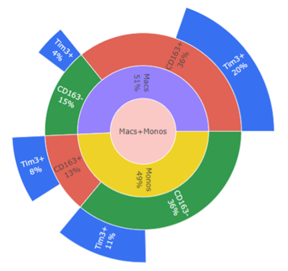
Biomarker Validation
Have a novel antibody or target you need to validate? IMCO provides an end-to-end pipeline, from wet lab optimization through quantitative correlation analysis. By benchmarking your candidate against gold standard assays with rigorous positive and negative controls, we ensure your biomarker delivers a signal that stands above the noise — giving you the confidence to move forward.

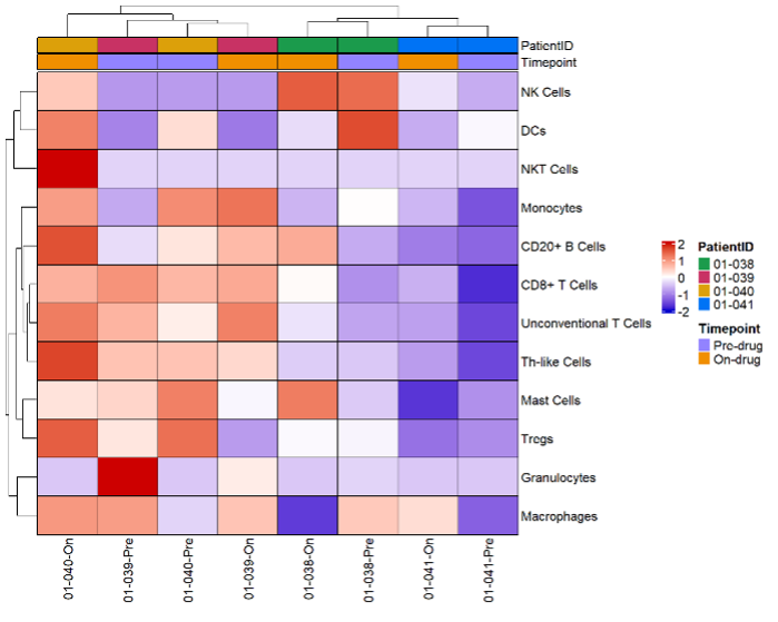
Exploratory Immune Profiling & Contexture Analysis
Starting with tissue samples and an undefined omic profile? We'll help you find your footing. IMCO delivers a comprehensive quantitative breakdown of the immune and cellular contexture of each sample, revealing hidden relationships and patterns across your dataset. Think of us as your research compass — orienting your findings, generating meaningful hypotheses, and empowering you to ask the right questions.
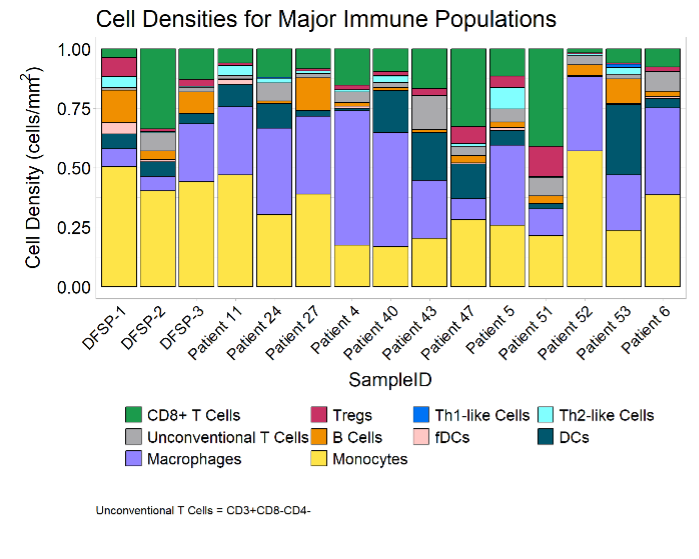
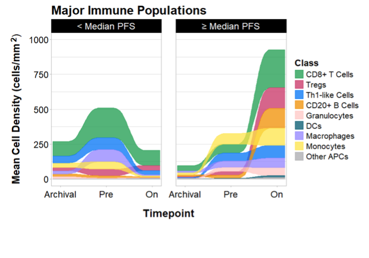
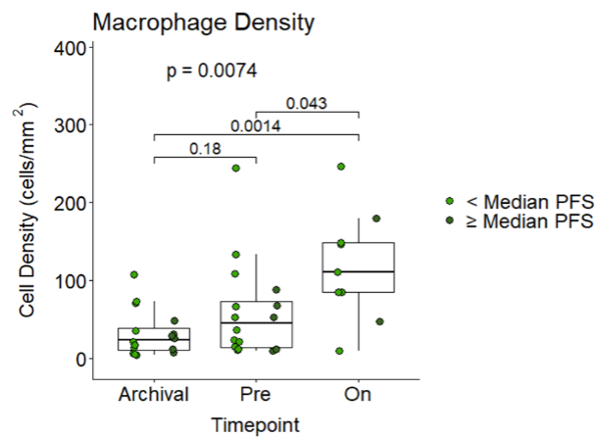
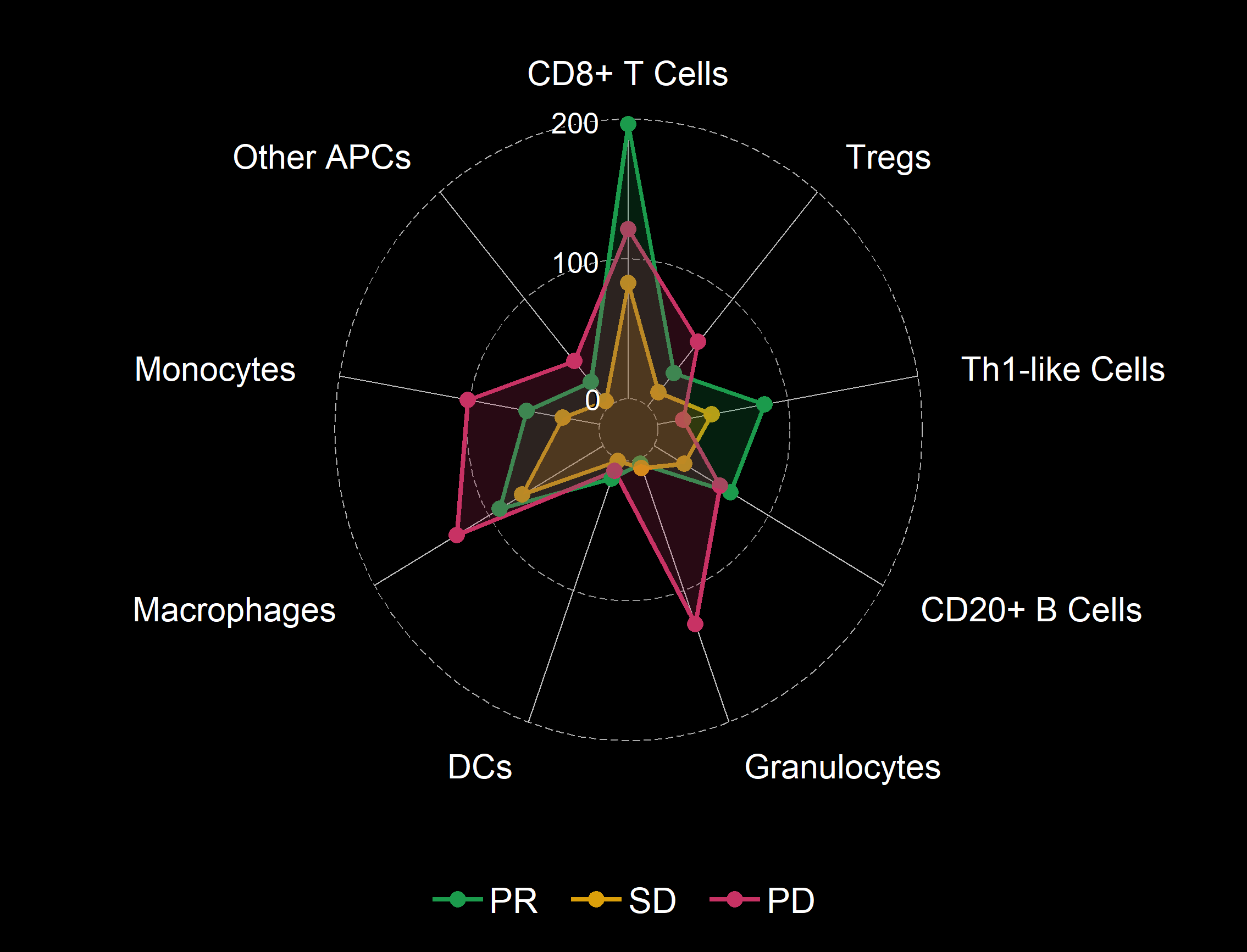
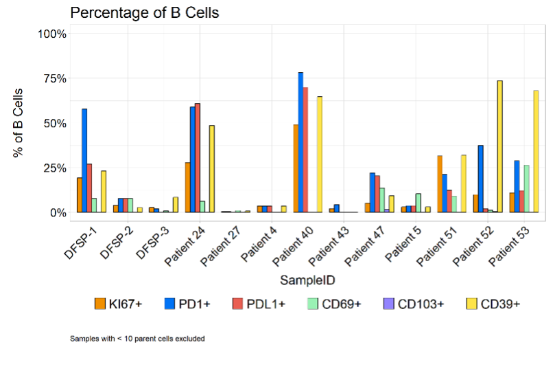
Clinical Cohort & Biomarker Analysis
For clinical trials or studies with well-defined research questions, IMCO offers rigorous cohort analysis tailored to your specific objectives. From longitudinal tracking across timepoints to responder vs. non-responder comparisons and multi-arm therapeutic evaluations, we deliver publication-ready visualizations, robust statistics, and spatial analysis. The result: clear, defensible evidence to support confident, data-driven decisions.
Clients receive:
QC reports
Statistical analysis summaries
Heatmaps
Volcano plots
PCA and UMAP visualizations
Differential expression analyses
Pathway enrichment reports
Publication-ready figures
Consultation and interpretation support
IMCO supports analysis of:
Olink proteomics
CycIF spatial biology
Mass cytometry (CyTOF)
Multiplex immunohistochemistry
RNA sequencing
Whole exome sequencing
Digital spatial profiling
Multi-omics datasets